Can Chloroplast Be Seen Under A Light Microscope . cells that have been fixed and stained can be studied in a conventional light. however, most organelles are not clearly visible by light microscopy, and those that can be seen (such as the nucleus,. Fluorescence microscopy uses fluorescent dyes (fluorophores),. viewed under a microscope, many green chloroplasts are visible. To view chloroplasts under the microscope, students can use toluidine blue stain to prepare a wet mount. being able to view chlorophyll under a microscope can help deepen your understanding of plant biology and. Each chloroplast contains neat stacks called grana. the nucleus and chloroplasts of eukaryotic cells can also be seen—however smaller organelles and viruses are beyond the limit of resolution of the light microscope (see.
from scopem.ethz.ch
the nucleus and chloroplasts of eukaryotic cells can also be seen—however smaller organelles and viruses are beyond the limit of resolution of the light microscope (see. To view chloroplasts under the microscope, students can use toluidine blue stain to prepare a wet mount. Fluorescence microscopy uses fluorescent dyes (fluorophores),. however, most organelles are not clearly visible by light microscopy, and those that can be seen (such as the nucleus,. Each chloroplast contains neat stacks called grana. cells that have been fixed and stained can be studied in a conventional light. viewed under a microscope, many green chloroplasts are visible. being able to view chlorophyll under a microscope can help deepen your understanding of plant biology and.
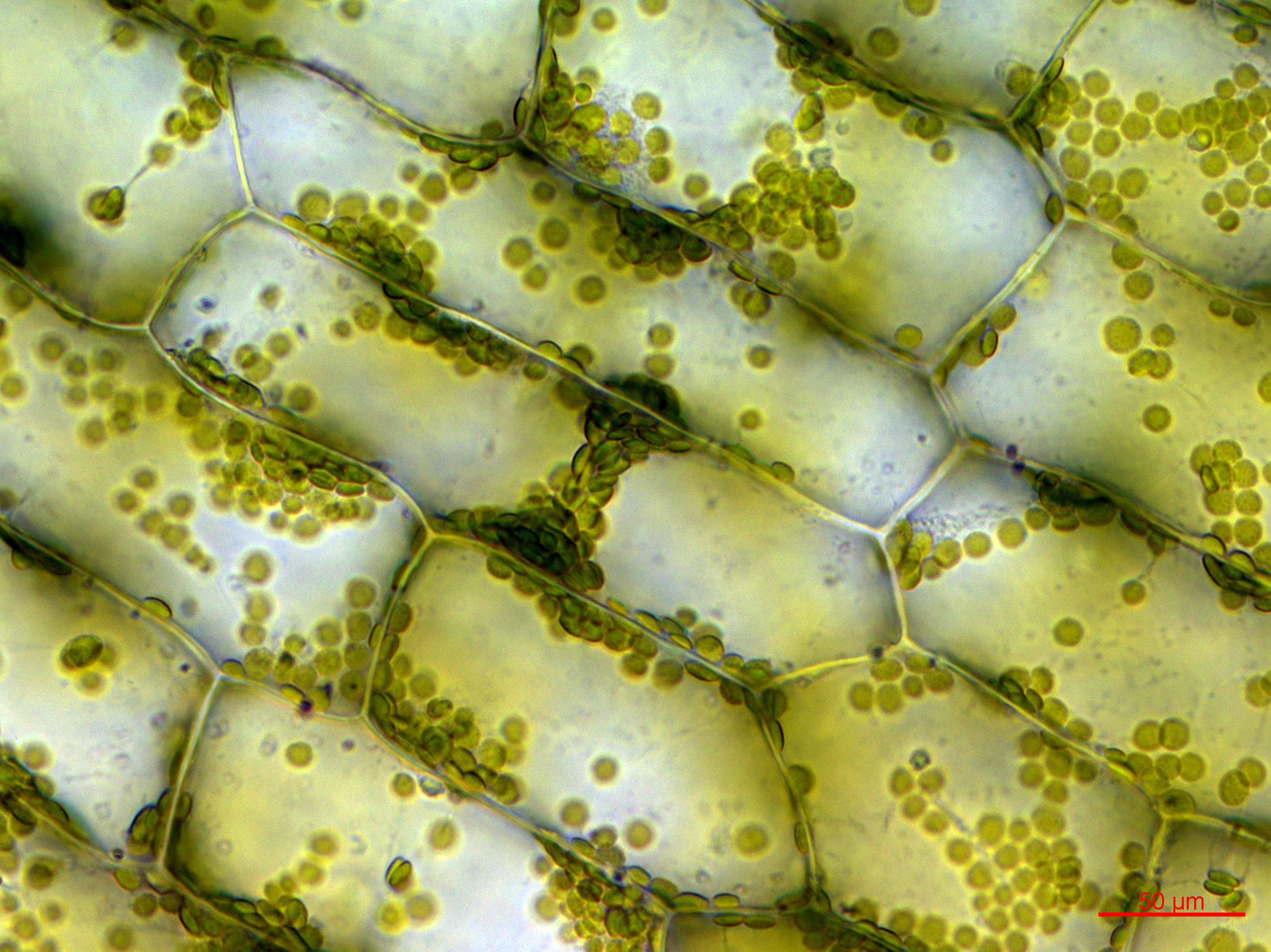
Chloroplast Scientific Center for Optical and Electron Microscopy
Can Chloroplast Be Seen Under A Light Microscope the nucleus and chloroplasts of eukaryotic cells can also be seen—however smaller organelles and viruses are beyond the limit of resolution of the light microscope (see. being able to view chlorophyll under a microscope can help deepen your understanding of plant biology and. cells that have been fixed and stained can be studied in a conventional light. however, most organelles are not clearly visible by light microscopy, and those that can be seen (such as the nucleus,. To view chloroplasts under the microscope, students can use toluidine blue stain to prepare a wet mount. viewed under a microscope, many green chloroplasts are visible. Each chloroplast contains neat stacks called grana. Fluorescence microscopy uses fluorescent dyes (fluorophores),. the nucleus and chloroplasts of eukaryotic cells can also be seen—however smaller organelles and viruses are beyond the limit of resolution of the light microscope (see.
From www.thoughtco.com
Chloroplast Function in Photosynthesis Can Chloroplast Be Seen Under A Light Microscope the nucleus and chloroplasts of eukaryotic cells can also be seen—however smaller organelles and viruses are beyond the limit of resolution of the light microscope (see. being able to view chlorophyll under a microscope can help deepen your understanding of plant biology and. however, most organelles are not clearly visible by light microscopy, and those that can. Can Chloroplast Be Seen Under A Light Microscope.
From www.linstitute.net
CIE A Level Biology复习笔记13.1.1 Chloroplasts翰林国际教育 Can Chloroplast Be Seen Under A Light Microscope viewed under a microscope, many green chloroplasts are visible. being able to view chlorophyll under a microscope can help deepen your understanding of plant biology and. the nucleus and chloroplasts of eukaryotic cells can also be seen—however smaller organelles and viruses are beyond the limit of resolution of the light microscope (see. cells that have been. Can Chloroplast Be Seen Under A Light Microscope.
From scihub.world
Can You See Chloroplast Under Light Microscope Can Chloroplast Be Seen Under A Light Microscope being able to view chlorophyll under a microscope can help deepen your understanding of plant biology and. To view chloroplasts under the microscope, students can use toluidine blue stain to prepare a wet mount. Fluorescence microscopy uses fluorescent dyes (fluorophores),. Each chloroplast contains neat stacks called grana. cells that have been fixed and stained can be studied in. Can Chloroplast Be Seen Under A Light Microscope.
From www.youtube.com
leaking chloroplast in algae under light microscope at 40x10x YouTube Can Chloroplast Be Seen Under A Light Microscope To view chloroplasts under the microscope, students can use toluidine blue stain to prepare a wet mount. cells that have been fixed and stained can be studied in a conventional light. viewed under a microscope, many green chloroplasts are visible. being able to view chlorophyll under a microscope can help deepen your understanding of plant biology and.. Can Chloroplast Be Seen Under A Light Microscope.
From lambdastudy.com
Microscopy LambdaStudy Can Chloroplast Be Seen Under A Light Microscope Each chloroplast contains neat stacks called grana. To view chloroplasts under the microscope, students can use toluidine blue stain to prepare a wet mount. cells that have been fixed and stained can be studied in a conventional light. however, most organelles are not clearly visible by light microscopy, and those that can be seen (such as the nucleus,.. Can Chloroplast Be Seen Under A Light Microscope.
From www.sciencefacts.net
Chloroplast Definition, Structure, Functions with Diagram Can Chloroplast Be Seen Under A Light Microscope To view chloroplasts under the microscope, students can use toluidine blue stain to prepare a wet mount. Fluorescence microscopy uses fluorescent dyes (fluorophores),. the nucleus and chloroplasts of eukaryotic cells can also be seen—however smaller organelles and viruses are beyond the limit of resolution of the light microscope (see. being able to view chlorophyll under a microscope can. Can Chloroplast Be Seen Under A Light Microscope.
From cartoondealer.com
Plant Cell With Chloroplast Under Microscope Stock Photo Can Chloroplast Be Seen Under A Light Microscope Fluorescence microscopy uses fluorescent dyes (fluorophores),. To view chloroplasts under the microscope, students can use toluidine blue stain to prepare a wet mount. being able to view chlorophyll under a microscope can help deepen your understanding of plant biology and. viewed under a microscope, many green chloroplasts are visible. Each chloroplast contains neat stacks called grana. cells. Can Chloroplast Be Seen Under A Light Microscope.
From scihub.world
Can You See Chloroplast Under Light Microscope Can Chloroplast Be Seen Under A Light Microscope the nucleus and chloroplasts of eukaryotic cells can also be seen—however smaller organelles and viruses are beyond the limit of resolution of the light microscope (see. viewed under a microscope, many green chloroplasts are visible. however, most organelles are not clearly visible by light microscopy, and those that can be seen (such as the nucleus,. cells. Can Chloroplast Be Seen Under A Light Microscope.
From mavink.com
Well Labelled Diagram Of Chloroplast Can Chloroplast Be Seen Under A Light Microscope viewed under a microscope, many green chloroplasts are visible. cells that have been fixed and stained can be studied in a conventional light. the nucleus and chloroplasts of eukaryotic cells can also be seen—however smaller organelles and viruses are beyond the limit of resolution of the light microscope (see. Each chloroplast contains neat stacks called grana. Fluorescence. Can Chloroplast Be Seen Under A Light Microscope.
From www.youtube.com
Moss leaf chloroplasts under microscope 1000x Ceratodon purpureus Can Chloroplast Be Seen Under A Light Microscope Fluorescence microscopy uses fluorescent dyes (fluorophores),. viewed under a microscope, many green chloroplasts are visible. being able to view chlorophyll under a microscope can help deepen your understanding of plant biology and. the nucleus and chloroplasts of eukaryotic cells can also be seen—however smaller organelles and viruses are beyond the limit of resolution of the light microscope. Can Chloroplast Be Seen Under A Light Microscope.
From www.alamy.com
Chloroplast TEM Stock Photo Alamy Can Chloroplast Be Seen Under A Light Microscope viewed under a microscope, many green chloroplasts are visible. the nucleus and chloroplasts of eukaryotic cells can also be seen—however smaller organelles and viruses are beyond the limit of resolution of the light microscope (see. being able to view chlorophyll under a microscope can help deepen your understanding of plant biology and. Each chloroplast contains neat stacks. Can Chloroplast Be Seen Under A Light Microscope.
From www.sciencephoto.com
Chloroplasts in Elodea cells, light micrograph Stock Image C038 Can Chloroplast Be Seen Under A Light Microscope To view chloroplasts under the microscope, students can use toluidine blue stain to prepare a wet mount. cells that have been fixed and stained can be studied in a conventional light. Each chloroplast contains neat stacks called grana. viewed under a microscope, many green chloroplasts are visible. Fluorescence microscopy uses fluorescent dyes (fluorophores),. the nucleus and chloroplasts. Can Chloroplast Be Seen Under A Light Microscope.
From www.researchgate.net
Induction, detection, and analyses of chloroplast photorelocation Can Chloroplast Be Seen Under A Light Microscope viewed under a microscope, many green chloroplasts are visible. however, most organelles are not clearly visible by light microscopy, and those that can be seen (such as the nucleus,. To view chloroplasts under the microscope, students can use toluidine blue stain to prepare a wet mount. being able to view chlorophyll under a microscope can help deepen. Can Chloroplast Be Seen Under A Light Microscope.
From www.kentfaith.co.uk
Can You See Chloroplast Under Light Microscope Can Chloroplast Be Seen Under A Light Microscope viewed under a microscope, many green chloroplasts are visible. the nucleus and chloroplasts of eukaryotic cells can also be seen—however smaller organelles and viruses are beyond the limit of resolution of the light microscope (see. To view chloroplasts under the microscope, students can use toluidine blue stain to prepare a wet mount. however, most organelles are not. Can Chloroplast Be Seen Under A Light Microscope.
From www.youtube.com
Chloroplast Moving YouTube Can Chloroplast Be Seen Under A Light Microscope being able to view chlorophyll under a microscope can help deepen your understanding of plant biology and. cells that have been fixed and stained can be studied in a conventional light. viewed under a microscope, many green chloroplasts are visible. the nucleus and chloroplasts of eukaryotic cells can also be seen—however smaller organelles and viruses are. Can Chloroplast Be Seen Under A Light Microscope.
From www.101diagrams.com
Diagram of Chloroplast 101 Diagrams Can Chloroplast Be Seen Under A Light Microscope being able to view chlorophyll under a microscope can help deepen your understanding of plant biology and. Fluorescence microscopy uses fluorescent dyes (fluorophores),. To view chloroplasts under the microscope, students can use toluidine blue stain to prepare a wet mount. the nucleus and chloroplasts of eukaryotic cells can also be seen—however smaller organelles and viruses are beyond the. Can Chloroplast Be Seen Under A Light Microscope.
From socratic.org
Can you expect to see mitochondria while using a light compound Can Chloroplast Be Seen Under A Light Microscope the nucleus and chloroplasts of eukaryotic cells can also be seen—however smaller organelles and viruses are beyond the limit of resolution of the light microscope (see. cells that have been fixed and stained can be studied in a conventional light. To view chloroplasts under the microscope, students can use toluidine blue stain to prepare a wet mount. Fluorescence. Can Chloroplast Be Seen Under A Light Microscope.
From www.myxxgirl.com
Chloroplast In Plant Cell Under Microscope Micropedia My XXX Hot Girl Can Chloroplast Be Seen Under A Light Microscope Each chloroplast contains neat stacks called grana. To view chloroplasts under the microscope, students can use toluidine blue stain to prepare a wet mount. Fluorescence microscopy uses fluorescent dyes (fluorophores),. cells that have been fixed and stained can be studied in a conventional light. being able to view chlorophyll under a microscope can help deepen your understanding of. Can Chloroplast Be Seen Under A Light Microscope.